
Pre-Med & Nursing: Anatomy Memory & Recall Protocols
Master gross anatomy, embryology, and histology in Nigerian medical and nursing schools. Learn the 4-pillar recall system to pass 2nd MBBS and OSPE exams with ease.

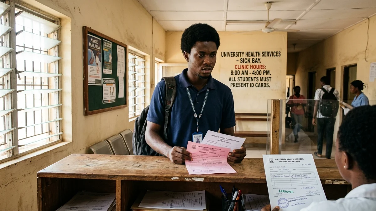
You step into the anatomy dissection hall at 8:00 AM.
The pungent, stinging smell of formalin hits your nostrils, making your eyes water behind your protective goggles. Stretched across twenty galvanized steel dissection tables are the cadavers your cohort will spend the next twelve months dissecting.
Beside you, a coursemate opens a massive, 1,200-page hardcover copy of Keith Moore’s Clinically Oriented Anatomy. Another student flips nervously through B.D. Chaurasia and Netter’s Atlas of Human Anatomy.
Your lecturer, a consultant anatomist, taps his forceps against a metallic tray:
*"In six months, you will face your 2nd Professional MBBS examinations. Anatomy does not accept approximations. If you cut the wrong nerve in a surgery, a patient is paralyzed. If you write the wrong origin or insertion on your script, you repeat the year. Begin dissecting the pectoral region."*
In Nigerian Colleges of Medicine and Faculties of Nursing—from CMUL (Idi-Araba) and UCH (Ibadan) to UNEC (Enugu) and ABU (Zaria)—the 2nd Professional MBBS / Nursing Examination is the most notorious academic bottleneck in tertiary education.
Regulated under strict standards set by the Medical and Dental Council of Nigeria (MDCN) and the Nursing and Midwifery Council of Nigeria (NMCN), pre-clinical students must memorize:
- Over **600 skeletal muscles** (origin, insertion, innervation, arterial supply, and actions).
- **12 pairs of cranial nerves** and their complex skull foramen exit trajectories.
- The intricate branches of the **brachial and lumbosacral plexuses**.
- Hundreds of histological slides, embryological folding stages, and clinical cross-sections.
Students who graduated top of their high school classes frequently drown in the sheer volume of Latin nomenclature. They highlight entire textbooks, stay awake until 4:00 AM breathing toxic formalin fumes, and still blank out during oral vivas and timed cadaver "spotter" exams.
Rote memorization fails because human working memory cannot hold thousands of disconnected facts without spatial anchoring.
Here is the 4-pillar cognitive retrieval system used by top medical and nursing scholars to memorize gross anatomy rapidly, retain clinical relations, and pass professional exams with distinctions.
1. The Cadaver-to-Script Spatial Anchoring Protocol
The biggest mistake pre-med and nursing students make is studying anatomy as flat, 2D textbook drawings.
In a written theory exam or in the cadaver hall, human anatomy exists in three-dimensional physical space. When an examiner asks you to describe the relations of the femoral triangle, reading a paragraph of text will fail you. You must construct a Mental Spatial Map:
- 2D Atlas Diagram] ➔ [3D Cadaver Dissection] ➔ [Blind Spatial Sketch from Memory
The 4 Compass Coordinates of Anatomical Relations:
Every anatomical structure in the human body has four mandatory relationship planes. When learning any organ, nerve, or artery, drill these four coordinates:
- **Anterior (Superficial):** What lies directly in front of it? (e.g., skin, fascia, specific muscular strap).
- **Posterior (Deep):** What sits directly behind it? (e.g., bone bed, deep tendon).
- **Medial:** What borders it toward the body's midline?
- **Lateral:** What borders it away from the midline?
When you memorize structures by their physical compass coordinates rather than memorizing sentences, you can mentally "walk through" the human body during an examination script write-up, effortlessly reproducing relations without memorizing word-for-word prose.
Just as laboratory procedures require structured write-up conventions, mastering practical histology and physiology reports demands rigorous formatting; see our guide on practical lab reports without penalties.
2. Image Occlusion & The Blind Sketch Drill
Stop highlighting Netter’s Atlas. Highlighting creates false recognition: you recognize the label "Musculocutaneous Nerve" because your eyes saw the line pointing to the coracobrachialis muscle.
In the exam hall, the arrows are removed.
How to Run Image Occlusion on Blank Paper:
- **The 3-Minute Mental Trace:** Look at an anatomical plate (e.g., the branches of the celiac trunk or the flexor compartment of the forearm) for three minutes.
- **Close the Atlas Completely:** Do not look back.
- **The Blind Sketch Drill:** Pull out a clean sheet of unlined A4 paper. Draw the structural skeleton using a blue ballpoint pen. Sketch the main arterial trunk and draw the branches from memory.
- **Label Every Terminal Branch:** Write out the names, nerve roots, and supplied muscles without looking at the textbook.
- **The Audit:** Compare your sketch with Netter in red ink. Any missing branch or misidentified relationship represents your exact cognitive blind spot.
If you can draw the brachial plexus blindly on a blank sheet of paper in under four minutes, you have mastered it permanently.
To structure these revision drills so you do not forget complex nerve pathways over twelve-week blocks, deploy our system on spaced repetition revision plan.
3. High-Yield Mnemonic & Story Architecture
Professional medical exams demand instant retrieval of complex sequences. Elite medical students do not rely on raw memory; they deploy associative clinical mnemonics.
3 Essential Mnemonics Every Pre-Med Must Know:
| Anatomical Region | Classic High-Yield Mnemonic | Clinical / Structural Reality |
|---|---|---|
| Carpal Bones (Wrist) | "Some Lovers Try Positions That They Can't Handle" | Scaphoid, Lunate, Triquetrum, Pisiform, Trapezium, Trapezoid, Capitate, Hamate |
| Bicipital Groove Muscles | "A Lady Between Two Majors" | Latissimus dorsi (the Lady) inserted between Pectoralis major and Teres major |
| Femoral Triangle Contents | "NAVEL (Lateral to Medial)" | Nerve, Artery, Vein, Empty space (canal), Lymphatics |
| Cranial Nerve Functions | "Some Say Marry Money, But My Brother Says Big Brains Matter More" | S = Sensory, M = Motor, B = Both (for CN I through XII) |
The Story Hook Technique:
For complex neuroanatomy tracts (such as the spinothalamic tract or corticospinal tract), turn the nerve fibers into a physical character:
- *Where was the neuron born?* (First order neuron in dorsal root ganglion).
- *Where does it cross the bridge?* (Decussates in the anterior white commissure of the spinal cord).
- *Where does it rest before the final journey?* (Synapses in the ventral posterolateral nucleus of the thalamus).
- *Where is its final destination?* (Primary somatosensory cortex, postcentral gyrus).
Turning cold neuroanatomy into an epic journey activates your brain’s episodic memory centers, preventing mental blocks.
4. The 60-Second OSPE / Spotter Sprint
In Nigerian pre-clinical exams, the Objective Structured Practical Examination (OSPE)—colloquially called "The Spotter"—strikes terror into candidates.
Twenty stations are arranged around the dissection laboratory. On each table rests a cadaveric limb, a wet organ pot, or a microscope with an unlabelled histology slide. A red or yellow pin marks a millimeter-thin structure.
A loud bell rings every 60 seconds. You must identify the structure, state its nerve supply, or write its embryological origin on your answer card before sprinting to the next table.
- **Bell Rings:** 00:00] ➔ [Identify Organ / Quadrant (10s)] ➔ [Differentiate Artery vs Nerve (15s)] ➔ [Write Full Latin Name (25s)] ➔ [Sanity Check (10s)
The Spotter Rule of Differentiation:
- **Feel the Structure (Elasticity):** In cadaveric specimens, arteries are thick-walled and rubbery; veins are collapsed and thin; nerves are solid, unyielding whitish cords.
- **Trace to Landmark Insertions:** Never guess a muscle in isolation. Trace the muscle fibers to their bony attachments (origin and insertion) before committing to a name.
5. Overcoming Formalin Fatigue & Mental Fog
Studying pre-clinical medicine is a physiological marathon. Constant exposure to formalin fumes in enclosed dissection halls causes mucosal irritation, severe eye strain, and physical fatigue.
When coupled with 5:00 AM wakeups and marathon clinical lectures, medical students often suffer from acute cognitive sluggishness.
If you find yourself reading the same anatomical paragraph four times without retaining a single Latin word, you are battling physiological brain fog; discover how to restore physical mental clarity in physical causes of student brain fog and fatigue.
The Pre-Med Daily Memory Checklist
Enforce this cognitive routine for every anatomy chapter:
- **Visualize in 3D:** Map every muscle and organ across its Anterior, Posterior, Medial, and Lateral coordinates.
- **Execute one Blind Sketch daily:** Draw plexuses, arterial trees, or organ cross-sections on unlined A4 paper.
- **Anchor complex sequences with mnemonics:** Use story hooks for cranial nerves, pathways, and fascial planes.
- **Run 60-second timed spotter flashcards:** Simulate the speed of professional OSPE examinations.
- **Protect 6 hours of sleep before dissection days:** Memory consolidation requires deep slow-wave neurological rest.
Secure Your Professional Medical & Nursing Distinctions
Surviving pre-clinical medicine and conquering 2nd MBBS requires more than raw willpower. You need an engineered study system that organizes massive volumes of complex data into crystal-clear recall.
The Academic Comeback Package
A comprehensive 7-part digital system engineered specifically for Nigerian university students ready to master technical courses, streamline study schedules, and produce top-tier examination results.
- Get Good at Hard Things — The cognitive framework to master anatomy, biochemistry, and complex clinical sciences
- How to Score High in Any Exam — Reverse-engineer marking rubrics, oral vivas, and professional exam criteria
- Focus Template — Maximize retention during 4-hour pre-clinical study blocks and eliminate mental fatigue
Frequently Asked Questions
Why is Gross Anatomy considered the hardest course in 2nd MBBS?
Gross Anatomy requires memorizing over 600 muscles (origins, insertions, nerve supplies, and actions), 206 bones, cranial nerve pathways, and complex 3D fascial planes. Unlike clinical courses based on reasoning, anatomy demands rapid, flawless nomenclature recall under extreme examination pressure.
How does the 'Image Occlusion' study method work for anatomy?
Image Occlusion involves taking an unlabeled anatomical illustration from Netter or Keith Moore, blocking out the label names with digital or physical boxes, and forcing yourself to name every pinned nerve, vessel, and muscle insertion from memory before revealing the answer.
How should medical students prepare for OSPE (Spotter) exams?
The Objective Structured Practical Examination (OSPE) allows only 60 seconds per station to identify pinned structures on cadavers, wet pots, and histology slides. Practice with timed 60-second flashcard drills, focusing on differentiating arteries from veins, nerve cords, and muscle bellies under pressure.
How many hours a day should a Nigerian pre-med student study?
Cognitive endurance in medicine is about deliberate intensity rather than marathon reading. 4 to 5 hours of high-focus, active spaced repetition with blind sketch drills outperforms 10 hours of passive, exhausted reading in the dissection hall.
What Did You Like Most About This Guide?
Share your favorite takeaway, a study technique you tested, or ask a question.